
Gray Matter Loss?
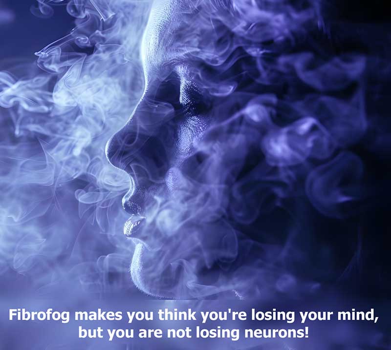
Multiple brain imaging studies report a loss of gray matter volume, mostly in pain processing areas such as the thalamus, prefrontal cortex, cingulate, amygdala, insula, and hippocampus. Yet, in fibromyalgia it does not appear to cause progressive or permanent structural changes to the neurons. In other words, fibromyalgia is not a disease like Alzheimer’s or Parkinson’s. So, what does the gray matter loss mean?
In most, if not all, of the areas showing reduced gray matter volume are regions with elevated glutamate levels. Although increased glutamate is toxic to the neurons, it’s also a sign that the astrocytes are activated and too sick to remove glutamate from the synapse. Once activated, the astrocytes (along with the microglia) change shape and become more compact. In addition, the activated glia may reduce blood flow and water content in the brain.
This chain of events may show up in brain imaging as a reduction in gray matter volume without any changes in the number of cells (neurons and glia combined). It is merely a volumetric change, sort of like squeezing a little water out of a sponge.
Another clue pointing to the glia as the reason for gray matter loss has to do with imaging studies looking at fibromyalgia patients taking opioids. Chronic use of opioids activates the glial cells, causing them to morph into a compact shape and reducing blood flow. Those patients on opioids had more gray matter volume loss than fibromyalgia patients not taking opioids … although both groups showed a loss compared to healthy people.
The spinal cord also contains gray matter, and the findings in the cord mimic that of the brain when it comes to assessing gray matter volumes. Despite a greater loss in gray matter volume, the patients on opioids showed a more normalized pain control … like that of healthy subjects. It’s as though taking opioids helps to intercept the impact of the incoming signals to the cord, while reducing the outgoing signals to the muscles.
Gray matter loss may simply reflect a change in the glia and the regional blood flow, rather than an actual loss of neurons. Longitudinal studies that follow patients over time, looking at glia activation and gray matter volumes, are needed to determine if these findings reflect glia “hot spots” in the brain.
The finding that opioids may help the pain-control system in fibromyalgia patients is both interesting and controversial. However, half of the opioid users were on tramadol, a mild opioid agonist that also raises CNS levels of serotonin and norepinephrine. But more importantly, these findings may explain why close to 75 percent of fibromyalgia patients insist that tramadol helps relieve their pain.