
Marker Maps Fibromyalgia Pain
Home | Treatment & Research News

A marker that maps fibromyalgia pain could validate what patients already know: their pain is widespread and real. One promising molecule is TSPO, short for translocator protein.
Many immune cells carry TSPO on their surface, especially when they respond to inflammation. This makes TSPO act like a flag that researchers can track while imaging the brain and other tissues. Marco Loggia, Ph.D., from Harvard Medical School, studies how TSPO may help detect inflammation-related pain. His work shows that TSPO signals can map painful areas in the brain, spinal cord, and possibly peripheral tissues.*
Neuroinflammation
To grasp why TSPO matters, it helps to first understand neuroinflammation. The term means ongoing inflammation in the brain and spinal cord. Glial cells drive much of this process. These support cells constantly watch for infection, injury, or other threats. In a healthy nervous system, glial cells respond quickly, help clean up the problem, and then return to their normal watchful state.
“Sometimes these immune responses become problematic,” says Loggia. “Activated glial cells continue unchecked even after the initial threat is resolved. They overstay their welcome and become harmful.” When this happens, glial cells may keep the nervous system inflamed. Researchers call this chronic neuroinflammation and have linked it to diseases such as Alzheimer’s, Parkinson’s, and schizophrenia.
Seeing TSPO Signals
Sampling brain tissue is too invasive. Instead, researchers can use PET, short for positron emission tomography, to image the brain. During PET imaging, doctors inject a small amount of radioactive tracer that attaches to TSPO and creates a signal on the scan.
Healthy brains show very little TSPO. But when neuroinflammation activates glial cells, TSPO levels rise and appear as “hot spots” on PET scans.
Loggia used PET to track TSPO in painful conditions linked to neuroinflammation, including fibromyalgia, low back pain, and migraines. These TSPO hot spots point to activated glial cells, which may help drive persistent pain. The location of the TSPO signal also tells researchers something important: it can show where the body hurts.
Mapping Pain Areas
Loggia’s first PET studies with a TSPO tracer focused on people with chronic back pain. Some patients showed a strong TSPO signal in the cortex, but others did not. Loggia suspected leg pain might explain the difference.
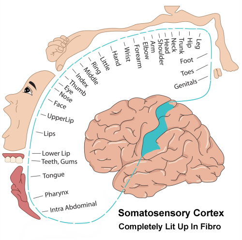
“Comparing patients with leg pain versus spinal pain, those with added leg pain showed a greater signal in the cortex,” says Loggia. He also measured each patient’s “fibromyalgia-like” score. The higher the score—the more painful body areas reported—the stronger the signal, especially in the somatosensory cortex.1
“The somatosensory cortex contains a full representation of the body,” says Loggia. “Different parts of this cortical region process sensory inputs from different body parts.” In low back pain patients, he says, “We saw a TSPO signal elevation in the cortex that matches the regional representation of back and leg pain.”
Migraines showed a similar pattern. The TSPO signal appeared in the cortical area that represents the face and head.
For fibromyalgia, Loggia says, “We should see the whole somatosensory cortex light up because patients report widespread pain. And that is what we see.”2 An AFSA-funded study later replicated this finding.
No Sex Differences
TSPO appears on immune cells, and the immune system can work differently in men and women. However, Loggia did not find sex differences in his low back pain studies.
Spinal Cord & DRGs
So far, these studies have focused on the brain. Can TSPO also detect pain-related inflammation in the spinal cord? Loggia showed that it can. In people with back pain, TSPO detected inflammation in the spinal cord and nearby structures, including the nerve roots and dorsal root ganglia, or DRGs.3
DRGs contain nerve cell bodies just before the nerves enter the spinal cord. Although DRGs sit outside the cord, TSPO-producing glial cells surround them. This matters for fibromyalgia because researchers suspect the DRGs may help amplify pain signals.
TSPO in Body Tissues
“Wouldn’t it be wonderful to use the same marker to image inflammation in the brain, spinal cord, and peripheral tissues like joints?” asks Loggia. Since many immune cells produce TSPO, it might work as a marker outside the nervous system too.
Loggia tested this idea in people with knee osteoarthritis. He compared PET scans from people with and without knee pain. Like the “hot spots” seen in the brain, TSPO signals appeared strongly in painful knees with arthritis. The signal also showed whether arthritis affected one or both knees.
Knee osteoarthritis is not fibromyalgia, but Loggia’s study found two points that may matter for chronic pain:
- Stronger TSPO signals matched higher levels of knee pain.
- TSPO signal strength also matched higher levels of several immune substances in the blood.
Together, these findings suggest that TSPO may help detect pain and inflammation in body tissues, although researchers still need more studies.
Tracking Treatment Response
If TSPO can detect pain and inflammation, it may also help researchers measure treatment response. Fibromyalgia studies often rely on symptom reports, which matter because patients know their pain best. But researchers also need objective tools that show whether a treatment changes pain-related inflammation.
Knee osteoarthritis offers one example. Total knee replacement removes the damaged joint, yet 30 percent of patients continue to have pain. Loggia found that TSPO signals in the brain before surgery helped predict who still had knee pain one year later.
The pre-surgery signal was stronger in patients who still had knee pain after one year. “This suggests that the TSPO signal we are measuring is not an epiphenomenon,” says Loggia. “It appears to have something to do with the establishment or maintenance of persistent pain.”
High-intensity signals in the sensory cortex of knee arthritis patients reflected the “fibromyalgia-like” nature of their pain. This pattern matters because fibromyalgia also involves widespread pain and increased TSPO signals in the sensory cortex. As researchers develop new fibromyalgia treatments, they might someday put them to the “TSPO test” to see whether they quiet pain-related inflammation.
Stay current on fibromyalgia treatment and research news.
Join AFSA’s free membership list for patient-friendly updates.
Future Markers
Loggia highlights TSPO as a marker for chronic pain and neuroinflammation. However, PET imaging with TSPO requires a radioactive tracer, so researchers continue to look for easier tools.
“Many groups around the world are looking at the concentration of brain metabolites believed to be linked to glial cell activity,” says Loggia. Researchers are studying these metabolites in fibromyalgia, but they still need to compare them with TSPO in the same patients. For now, Loggia concludes, “The use of TSPO to image peripheral and central nervous system inflammation (and pain) is opening new doors.”
Mapping Pain Beyond the Brain
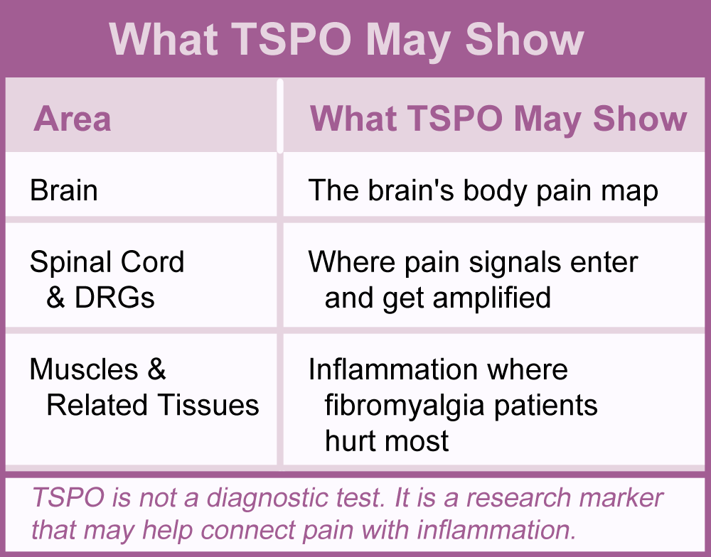
AFSA recently funded Loggia to use TSPO imaging in a new way. His team will look for inflammation outside the brain in areas where fibromyalgia patients hurt most, such as their muscles. The goal is not to turn whole-body PET into a diagnostic test. Instead, this study will test whether painful body areas also carry stronger TSPO signals. If they do, this fibromyalgia pain marker could help link immune activity and inflammation to patients’ pain.
TSPO appears on many immune cells, not just glial cells. When inflammation activates these cells, they may produce more TSPO. If the study finds stronger TSPO signals in painful muscles or other tissues, it could help validate the body-wide pain patients describe. It could also guide future treatment research.
Help move fibromyalgia research forward.
AFSA-funded studies like Dr. Loggia’s TSPO project may help validate pain patients feel but others cannot see. Your donation helps support more research toward better answers and treatments.
More About Glial Cells
While Loggia describes TSPO as a pain-mapping marker on immune cells, including glial cells, it helps to know what these cells are doing in fibromyalgia.
Glial Cells in Your Brain – These cells can be your neuron’s best friend, but research shows this is not the case for fibromyalgia patients. Find out what they are doing and what can be done to get them to behave.
Glia Around Your DRG – These cells can control your dorsal root ganglia (DRG) so the signal entering your spinal cord is amplified. Research shows your glia are under attack in fibromyalgia.
Symptoms | Medications | Alternative Therapies | Muscle Pain Relief | Fibro Friendly Exercises
1. Alshelh Z, Loggia ML, et al. Brain 145(3):1098-1110, 2022. Free Article
2. Albrecht DS, Loggia ML, et al. Brain Behav Immun 75:72-83, 2019. Free Article
3. Albrecht DS, Loggia ML, et al. Pain 159(5)968-977, 2018. Free Article
* Neuroinflammation: Does it have a role in human chronic pain? by Marco Loggia, Ph.D., presented at the International Association for the Study of Pain (IASP) World Congress, August 5, 2024.